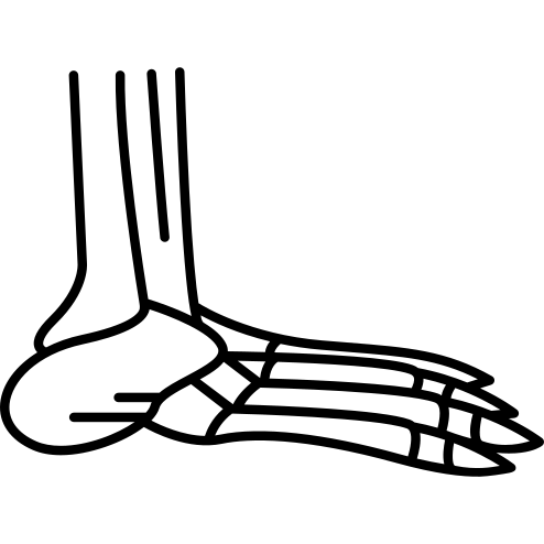
This detailed illustration showcases the various bones of the human foot, including the tarsals, metatarsals, and phalanges. The diagram serves as an educational tool for students, medical professionals, and anatomy enthusiasts, providing a clear visual representation of the foot's skeletal structure. It highlights the intricate arrangement of bones that support weight, enable movement, and absorb shock. This image is ideal for use in medical textbooks, anatomy courses, physical therapy materials, and presentations focused on human anatomy or podiatry. Its clarity and precision make it an excellent resource for understanding foot mechanics and common conditions related to foot health.


Part of the
Human Body Parts SVG collection,

in Healthcare.
Download the Anatomical Foot Bones Diagram vector as a ready-to-edit .svg file —
it scales to any size without losing quality and can be recoloured in any
vector editor or straight in CSS.